Chromosome Identification
Chromosome isolation and microscopic observation forms the basis of cytogenetics and is the primary method by which clinicians detect chromosomal abnormalities in humans. A karyotype is the number and appearance of chromosomes, and includes their length, banding pattern, and centromere position. To obtain a view of an individual’s karyotype, cytologists photograph the chromosomes and then cut and paste each chromosome into a chart, or karyogram. Another name is an ideogram (Figure).
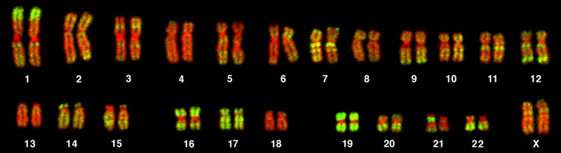
In a given species, we can identify chromosomes by their number, size, centromere position, and banding pattern. In a human karyotype, autosomes or “body chromosomes” (all of the non–sex chromosomes) are generally organized in approximate order of size from largest (chromosome 1) to smallest (chromosome 22). The X and Y chromosomes are not autosomes. However, chromosome 21 is actually shorter than chromosome 22. Researchers discovered this after naming Down syndrome as trisomy 21, reflecting how this disease results from possessing one extra chromosome 21 (three total). Not wanting to change the name of this important disease, scientists retained the numbering of chromosome 21 despite describing it having the shortest set of chromosomes. We may designate the chromosome “arms” projecting from either end of the centromere as short or long, depending on their relative lengths. We abbreviate the short arm p (for “petite”); whereas, we abbreviate the long arm q (because it follows “p” alphabetically). Numbers further subdivide and denote each arm. Using this naming system, we can describe chromosome locations consistently in the scientific literature.
Career Connection
Geneticists Use Karyograms to Identify Chromosomal AberrationsAlthough we refer to Mendel as the “father of modern genetics,” he performed his experiments with none of the tools that the geneticists of today routinely employ. One such powerful cytological technique is karyotyping, a method in which geneticists can identify traits characterized by chromosomal abnormalities from a single cell. To observe an individual’s karyotype, a geneticist first collects a person’s cells (like white blood cells) from a blood sample or other tissue. In the laboratory, he or she stimulates the isolated cells to begin actively dividing. The geneticist then applies the chemical colchicine to cells to arrest condensed chromosomes in metaphase. The geneticist then induces swelling in the cells using a hypotonic solution so the chromosomes spread apart. Finally, the geneticist preserves the sample in a fixative and applies it to a slide.
The geneticist then stains chromosomes with one of several dyes to better visualize each chromosome pair's distinct and reproducible banding patterns. Following staining, the geneticist views the chromosomes using bright-field microscopy. A common stain choice is the Giemsa stain. Giemsa staining results in approximately 400–800 bands (of tightly coiled DNA and condensed proteins) arranged along all 23 chromosome pairs. An experienced geneticist can identify each band. In addition to the banding patterns, geneticists further identify chromosomes on the basis of size and centromere location. To obtain the classic depiction of the karyotype in which homologous chromosome pairs align in numerical order from longest to shortest, the geneticist obtains a digital image, identifies each chromosome, and manually arranges the chromosomes into this pattern (Figure).
At its most basic, the karyogram may reveal genetic abnormalities in which an individual has too many or too few chromosomes per cell. Examples of this are Down Syndrome, which one identifies by a third copy of chromosome 21, and Turner Syndrome, which has the presence of only one X chromosome in women instead of the normal two characterizes. Geneticists can also identify large DNA deletions or insertions. For instance, geneticists can identify Jacobsen Syndrome—which involves distinctive facial features as well as heart and bleeding defects—by a deletion on chromosome 11. Finally, the karyotype can pinpoint translocations, which occur when a segment of genetic material breaks from one chromosome and reattaches to another chromosome or to a different part of the same chromosome. Translocations are implicated in certain cancers, including chronic myelogenous leukemia.
During Mendel’s lifetime, inheritance was an abstract concept that one could only infer by performing crosses and observing the traits that offspring expressed. By observing a karyogram, today’s geneticists can actually visualize an individual's chromosomal composition to confirm or predict genetic abnormalities in offspring, even before birth.